
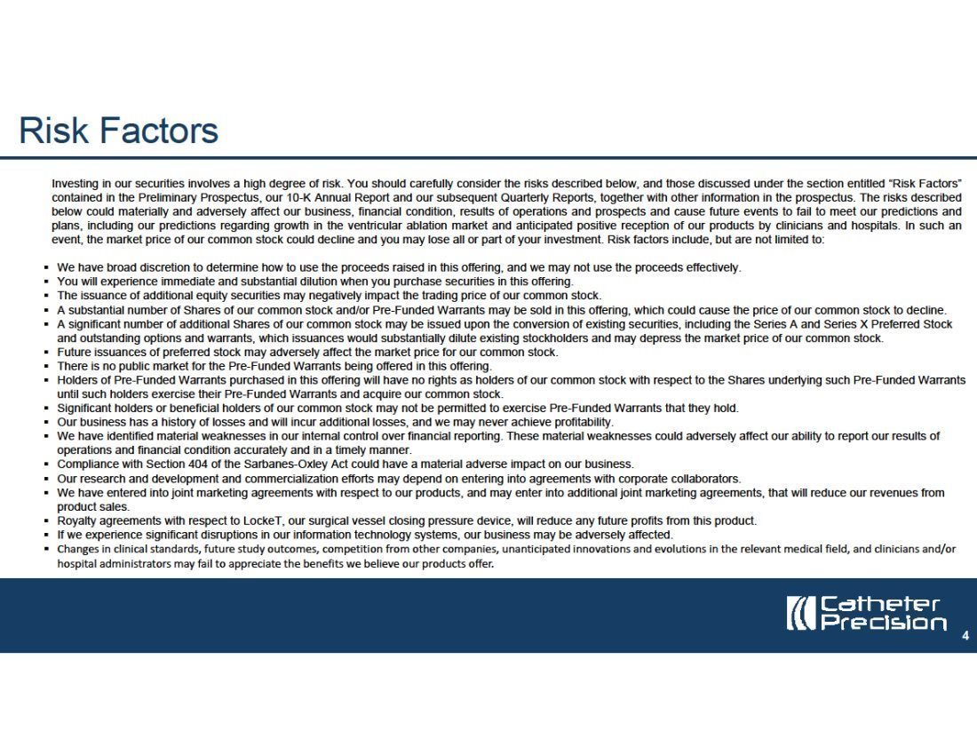


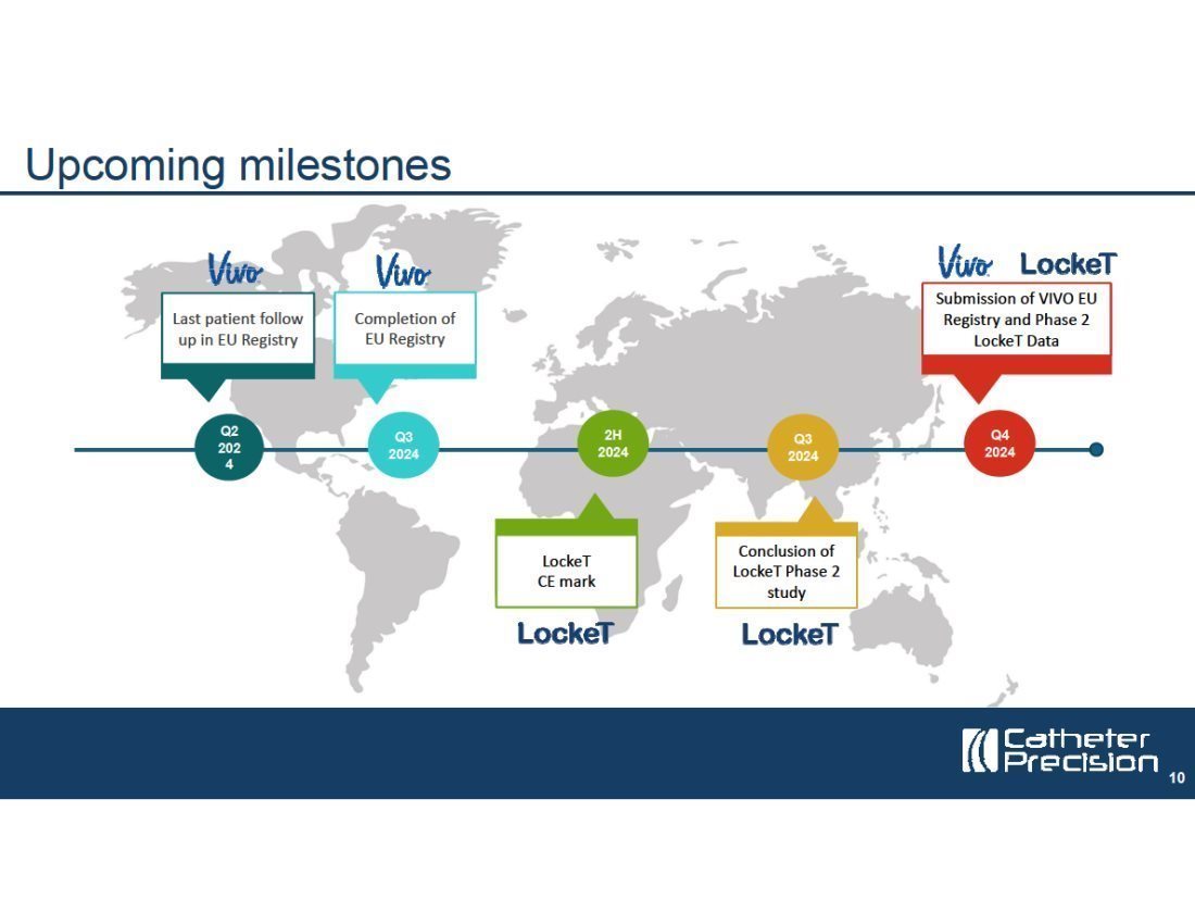


Appendix A
Description of embedded animation
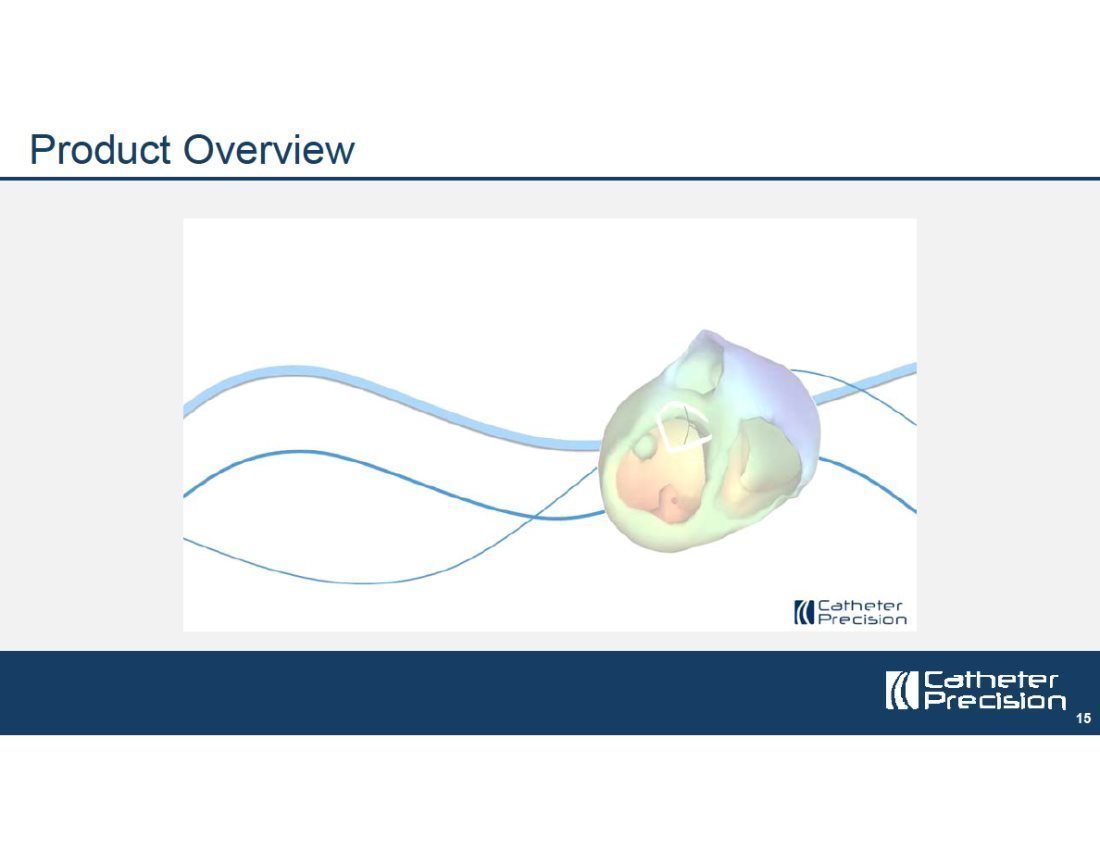
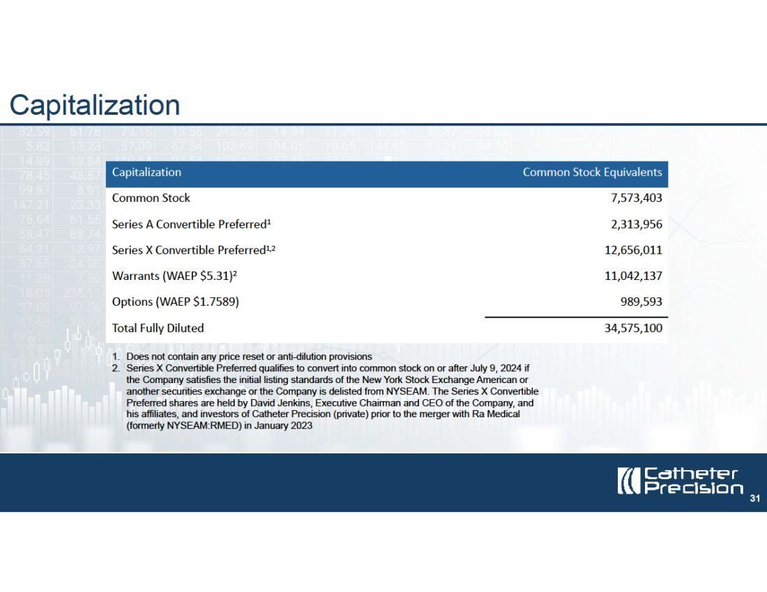
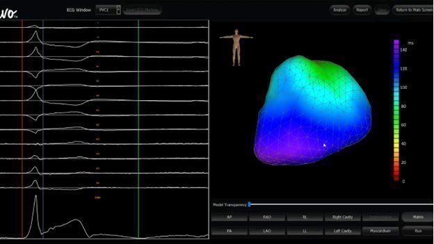
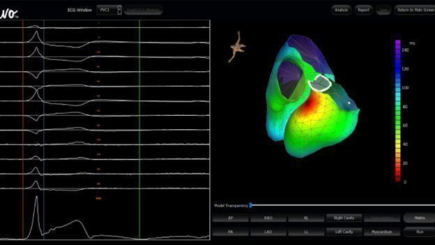
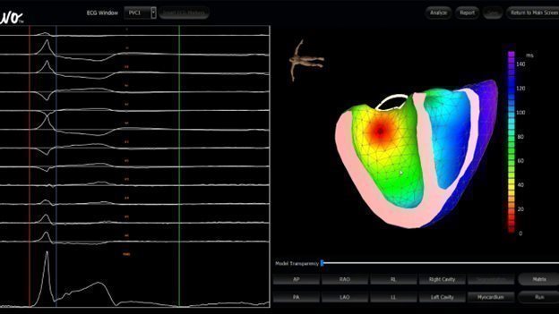
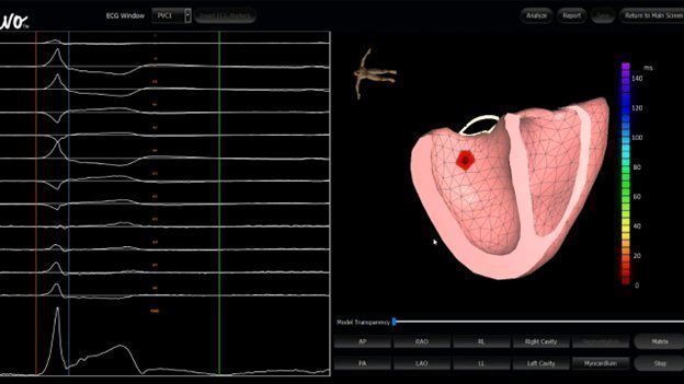
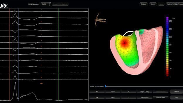
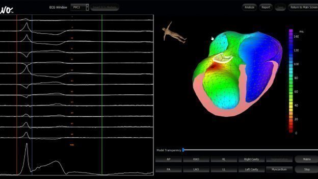
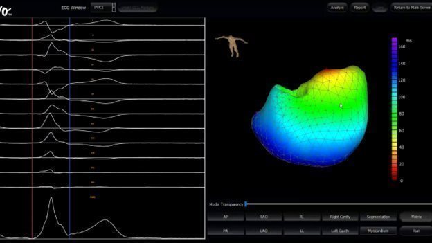
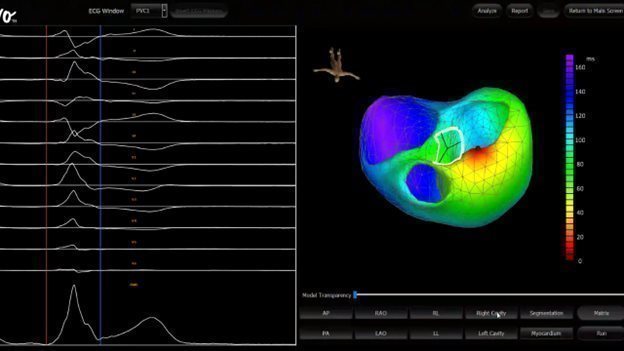
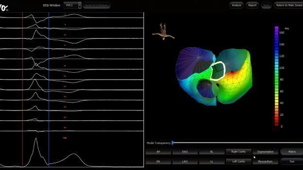
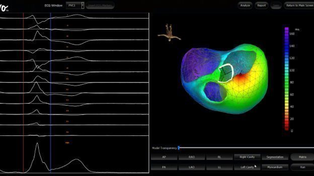
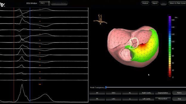
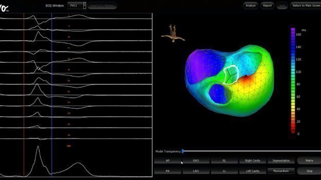
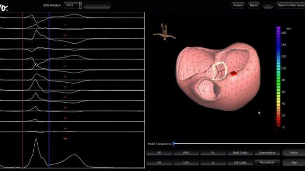
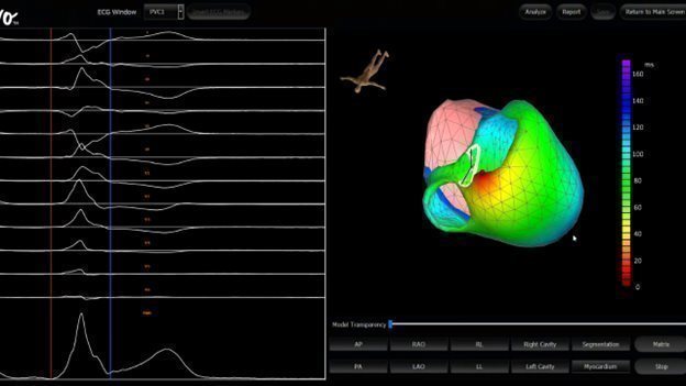
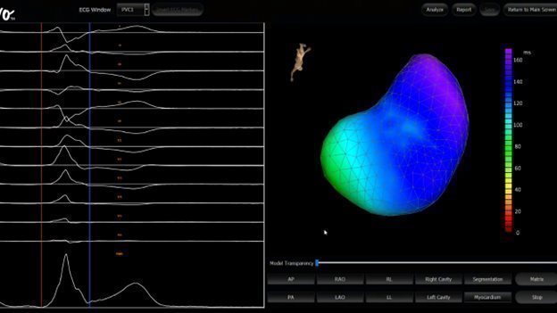
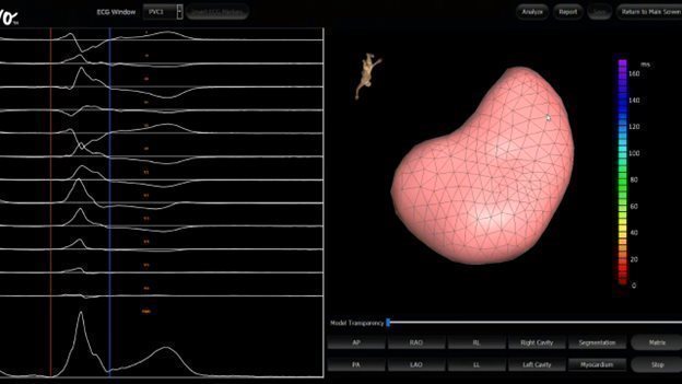
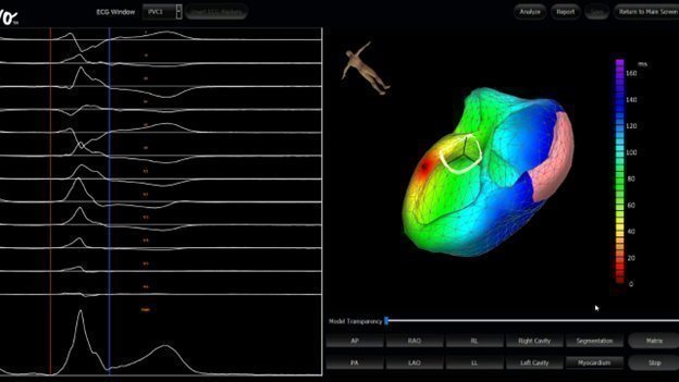
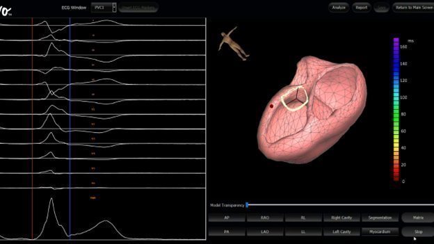
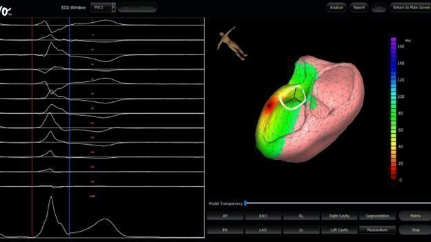
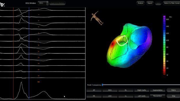
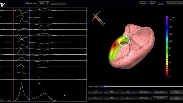
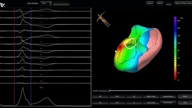
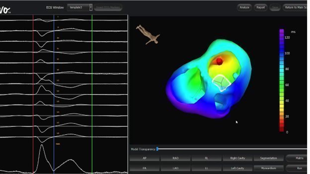
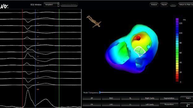
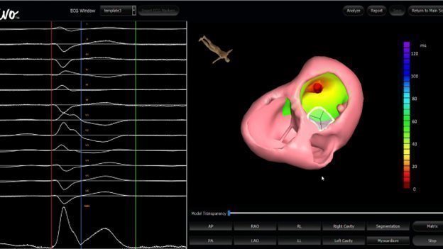
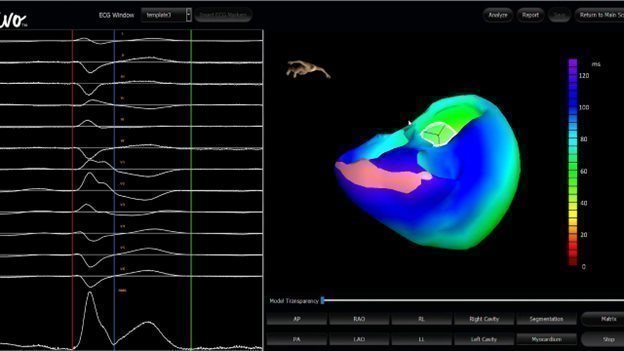
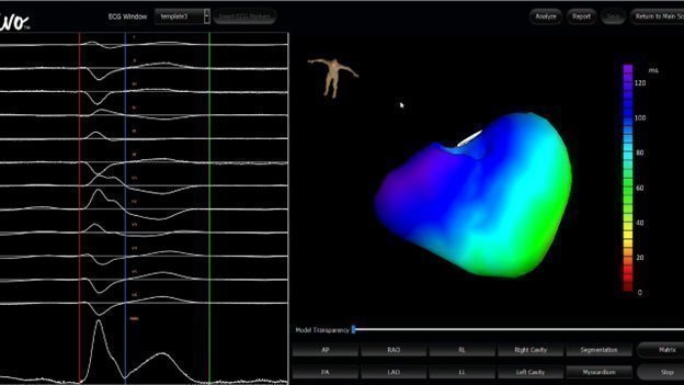
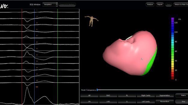
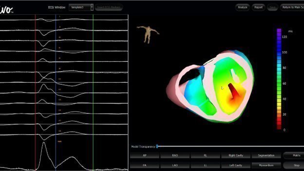
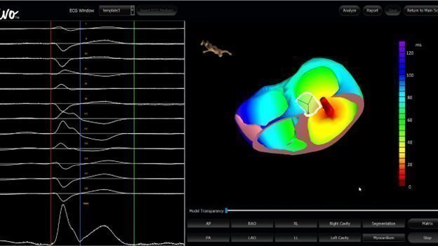
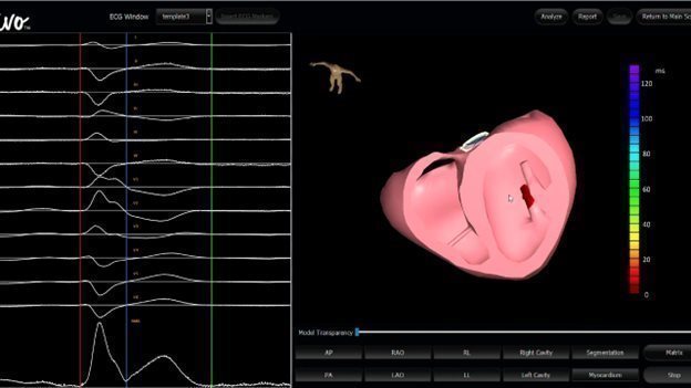
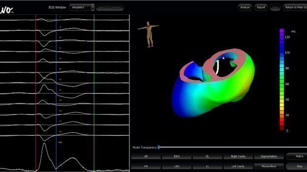
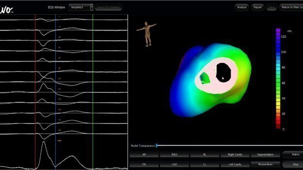
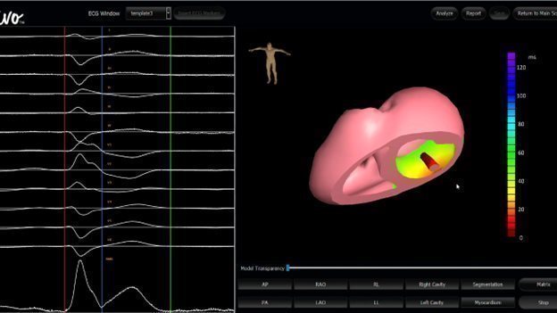
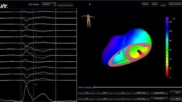
Slide 15 of the Presentation includes an embedded animation, without audio, that illustrates how VIVO works through a simulation of the user interface, including a 3D model of a patient, the patient’s heart, an EKG, and a user panel. Set forth below are representative graphic images from the embedded animation:


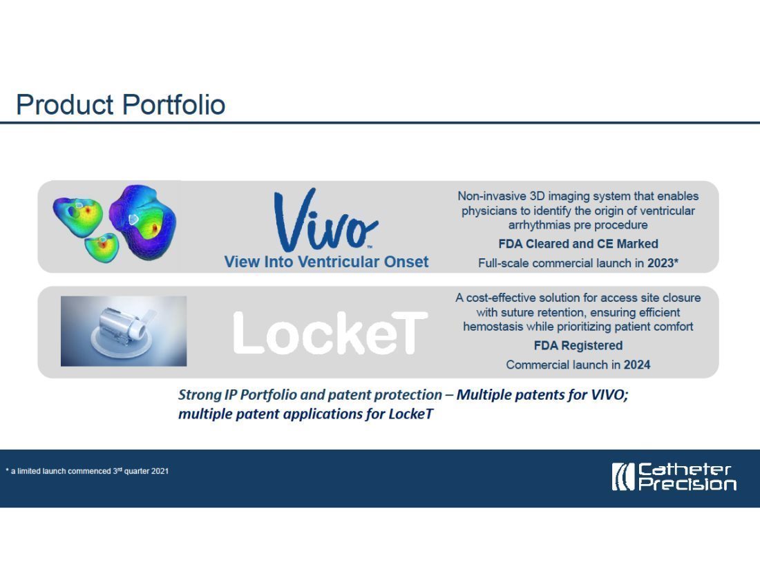
Appendix B
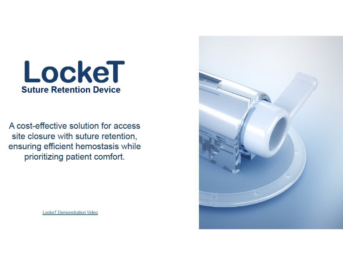
Hyperlinked LockeT Demonstration Video (Slide 20)
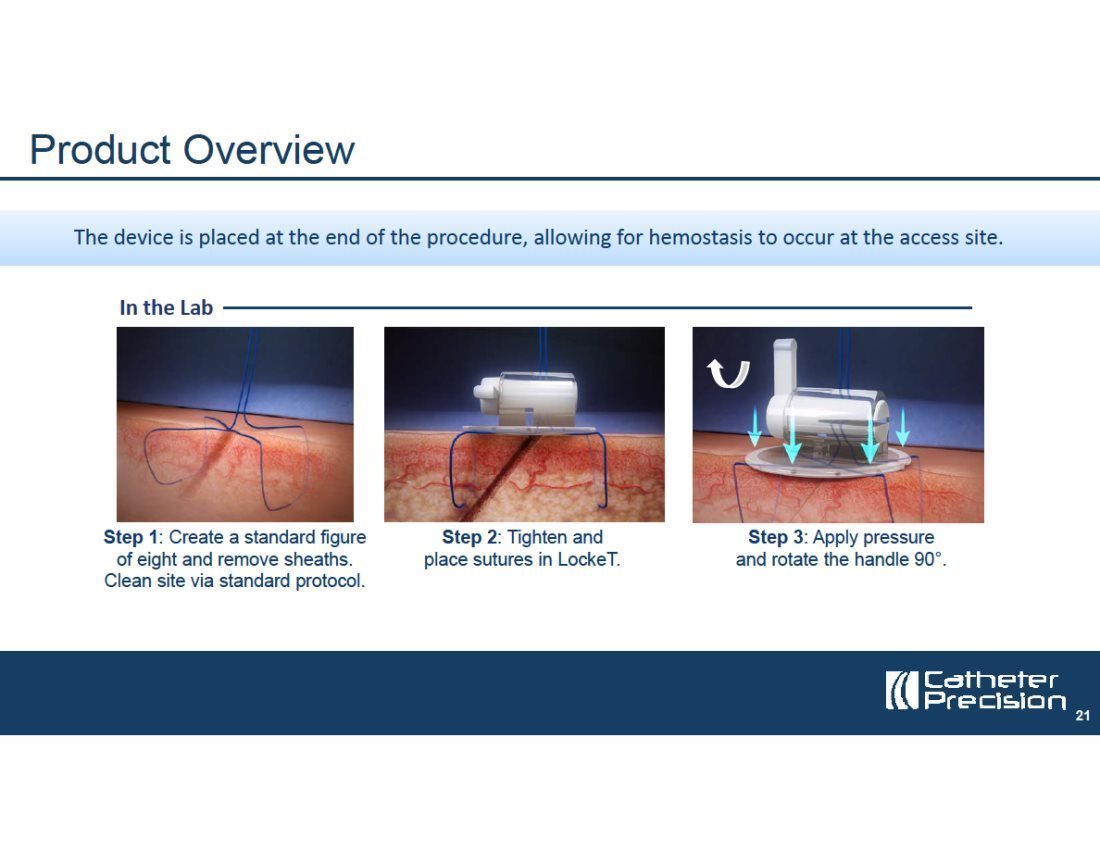
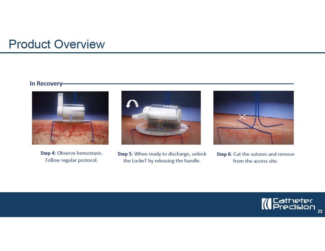
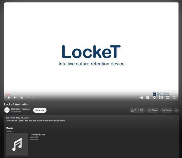
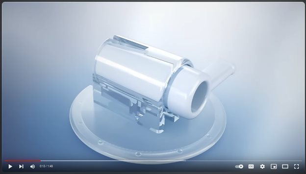
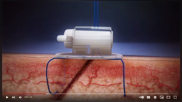
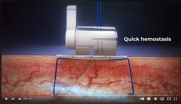
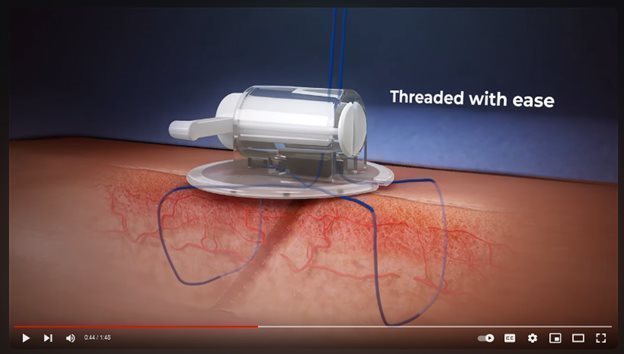
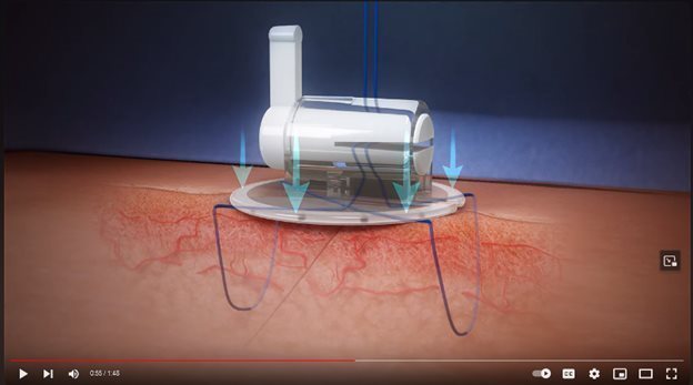
A hyperlink is provided to an animated video which illustrates the functioning and use of LockeT, hosted on the YouTube platform at www.youtube.com/watch?v=SUQZbqAnGhQ. A transcript of the video follows representative graphic images from the embedded video below.
| Transcript | |
|
|
|
| 0:00 | [Music] |
|
|
|
| 0:04 | Achieving hemostasis following large |
|
|
|
| 0:07 | bore femoral vein access remains a |
|
|
|
| 0:11 | challenge. Catheter Precision's LockeT |
|
|
|
| 0:14 | suture retention device features a |
|
|
|
| 0:16 | simple design, offering an easy to use, |
|
|
|
| 0:18 | comfortable and cost-effective solution |
|
|
|
| 0:20 | for hemostasis in small and large bore |
|
|
|
| 0:23 | venous access sites. LockeT helps |
|
|
|
| 0:26 | clinicians quickly achieve hemostasis |
|
|
|
| 0:31 | and remove sutures efficiently to help |
|
|
|
| 0:33 | patients heal faster and hospitals |
|
|
|
| 0:35 | achieve same day |
| 0:38 | discharge following standard figure-of-eight |
|
|
|
| 0:40 | suturing closure techniques. LockeT is |
|
|
|
| 0:43 | threaded onto sutures with ease. After |
|
|
|
| 0:45 | pulling the excess suture into the |
|
|
|
| 0:47 | slotted spindle, the handle is turned to |
|
|
|
| 0:50 | lock the thread and apply external |
|
|
|
| 0:52 | pressure to the venous access site, |
|
|
|
| 0:54 | rapidly achieving |
|
|
|
| 0:55 | hemostasis. Removal is easy: simply open |
|
|
|
| 0:58 | the stopcock valve, |
|
|
|
| 1:02 | lift and remove the |
|
|
|
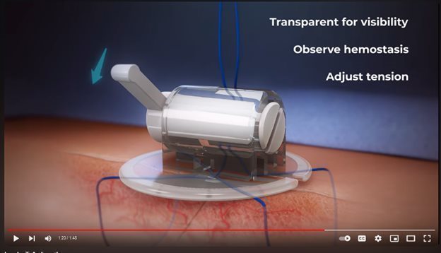
| 1:07 | suture. LockeT's unique transparent |
|
|
|
| 1:09 | design allows visibility to the catheter |
|
|
|
| 1:12 | access site, making it simple to observe |
|
|
|
| 1:14 | hemostasis and remove. The design of the |
|
|
|
| 1:17 | handle enables easy adjustment of suture |
|
|
|
| 1:21 | [Music] |
|
|
|
| 1:22 | tension. LockeT’s simple application and |
|
|
|
| 1:25 | removal equals time and cost savings, |
|
|
|
| 1:28 | obviating the need for manual |
|
|
|
| 1:30 | compression and offering a more |
|
|
|
| 1:31 | economical option compared to other |
|
|
|
| 1:33 | devices. Patients benefit from the |
|
|
|
| 1:35 | enhanced comfort and same day discharge. |
|
|
|
| 1:39 | [Music] |
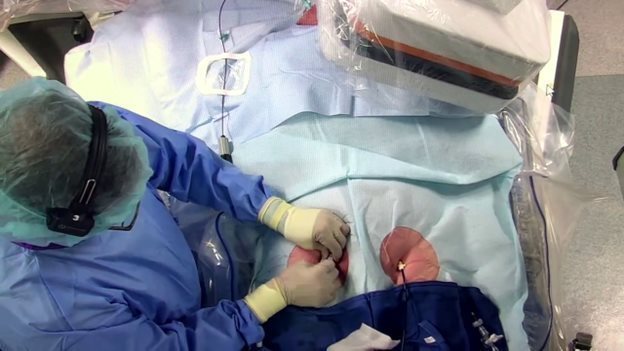
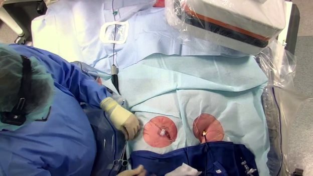
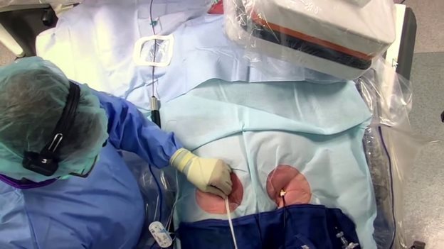
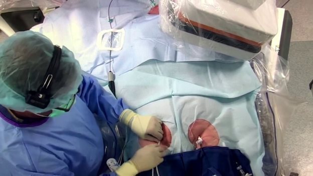
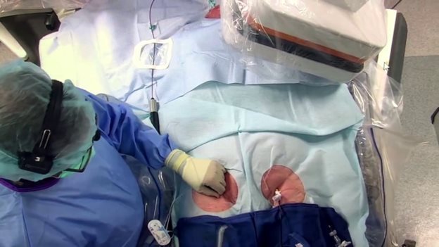
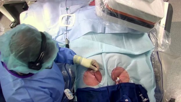
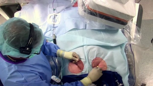
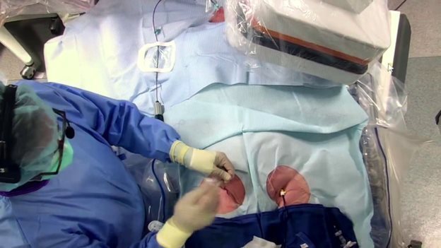
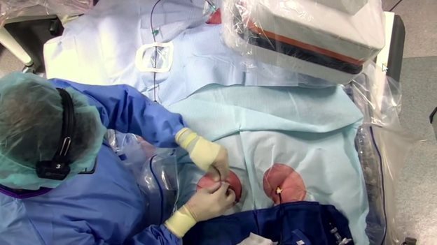
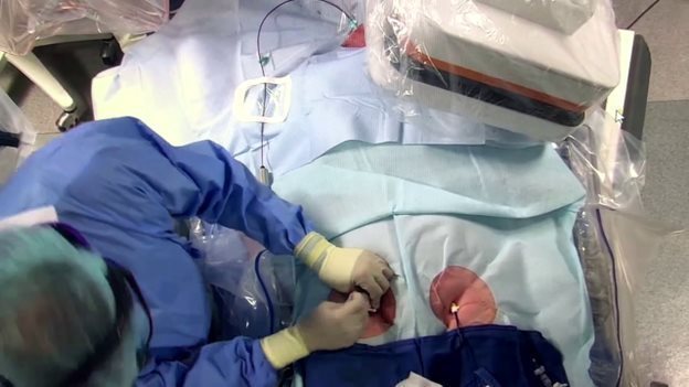
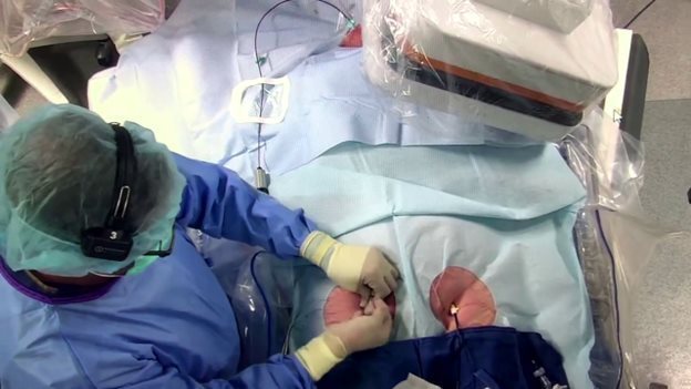
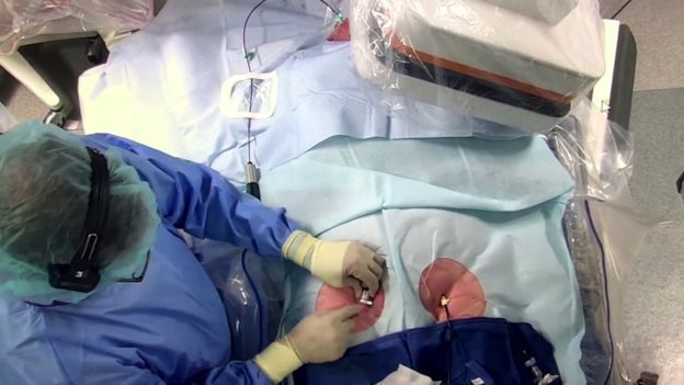
Appendix C
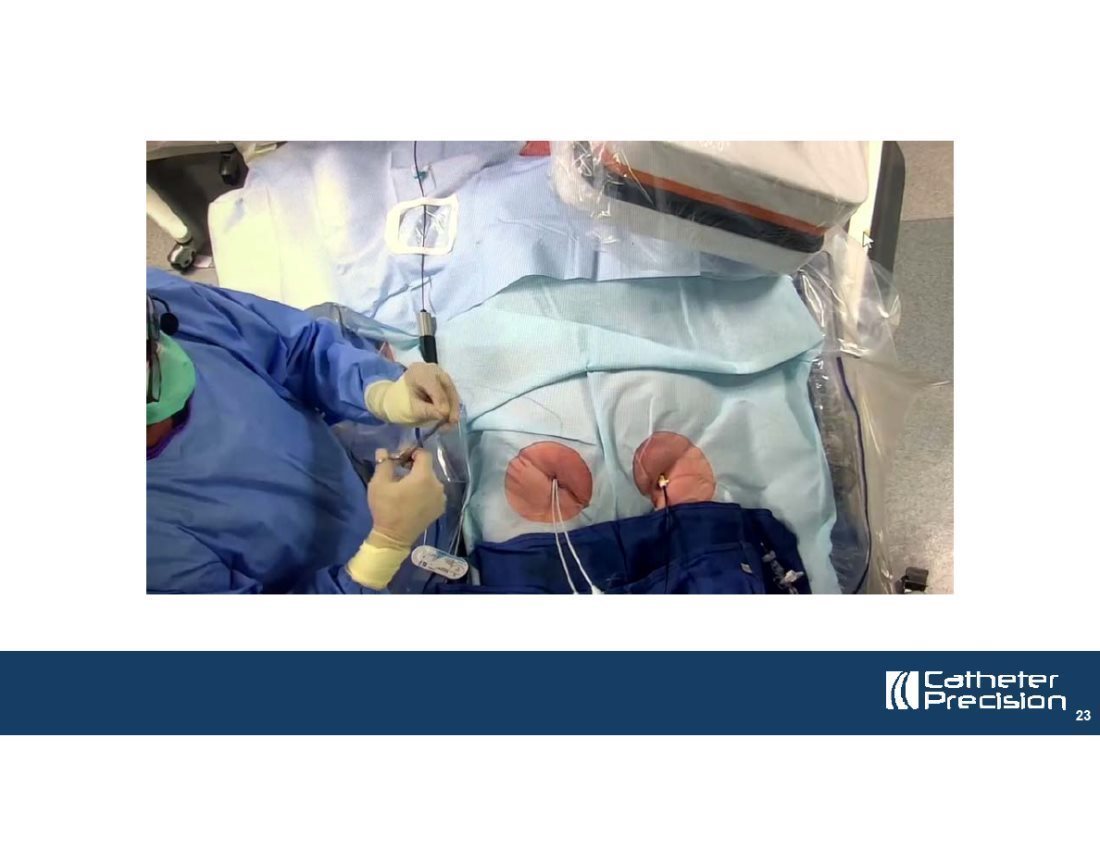
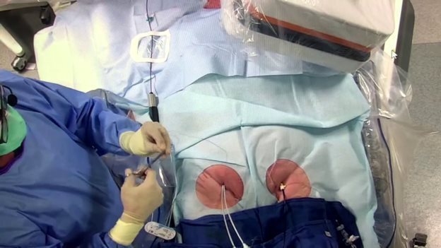
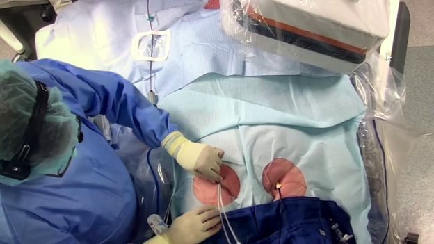
Description of embedded video (Slide 23)
An embedded live action video, without audio, in which a surgeon demonstrates the use of LockeT during access site closure following an ablation procedure. Set forth below are representative graphic images from the embedded video:


+